
Bipasha Kundu
Research Assistant at Chester F. Carlson Center for Imaging Science, Rochester Institute of Technology
- Rochester, NY
- Rochester Institute of Technology
- Google Scholar
- Github
Research Projects
Exploring the performance and robustness of Natural Domain Vision Language models for Segmenting Medical Image
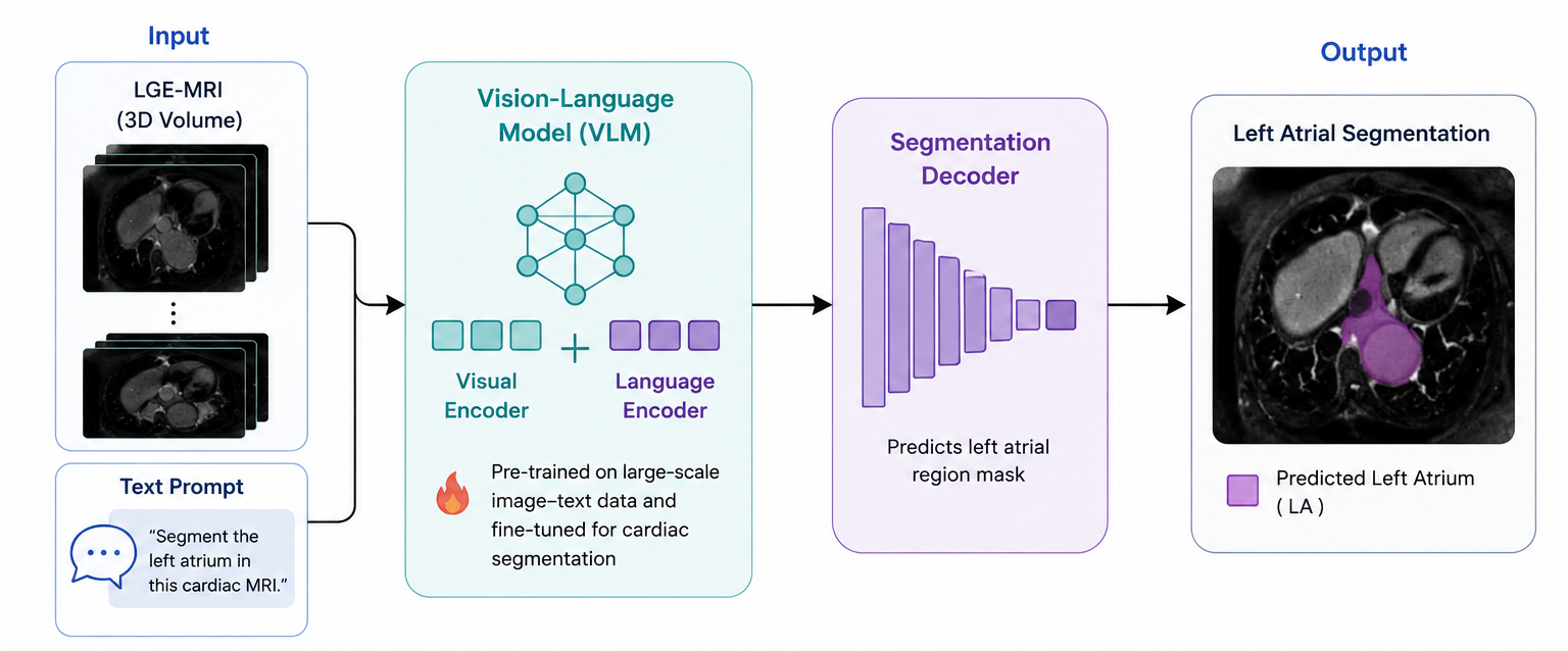 Project description coming soon.
Exploring the performance and robustness of Vision Language models for Image Quality Assessment
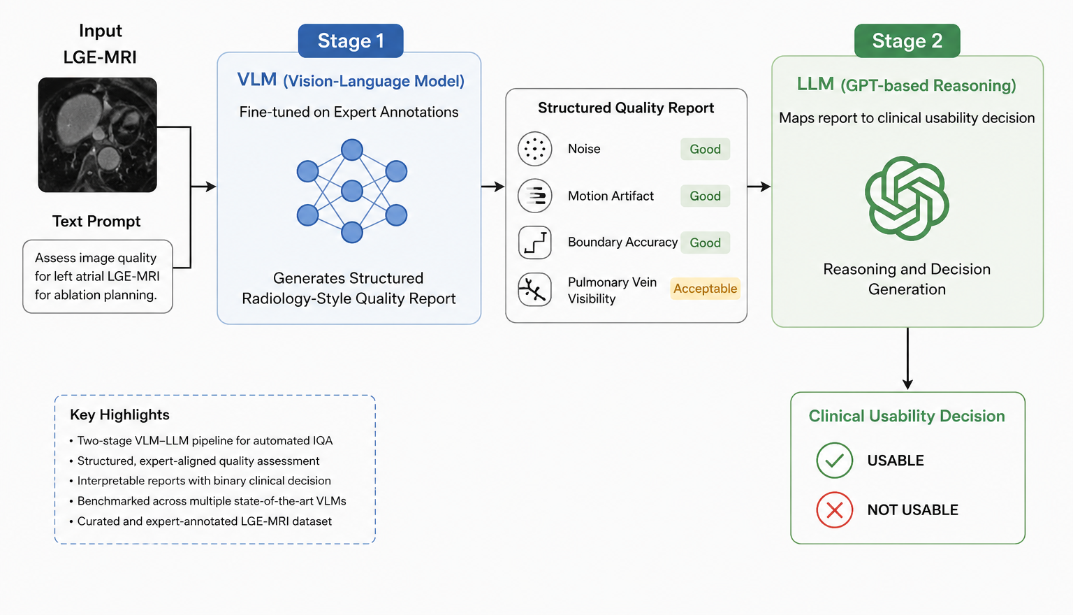 Developed a two-stage VLM-LLM framework for automated, clinically grounded image quality assessment of left atrial LGE-MRI scans to support ablation planning in atrial fibrillation patients. A fine-tuned VLM generates structured radiology-style quality reports across four expert-defined criteria, which are then mapped by a GPT-based reasoning module to a binary clinical usability decision. Benchmarked four state-of-the-art VLM architectures on a curated and expert-annotated MRI data.
Exploring the robustness of Vision Foundation Models for Left Atrium Segmentation from Cardiac MR Images
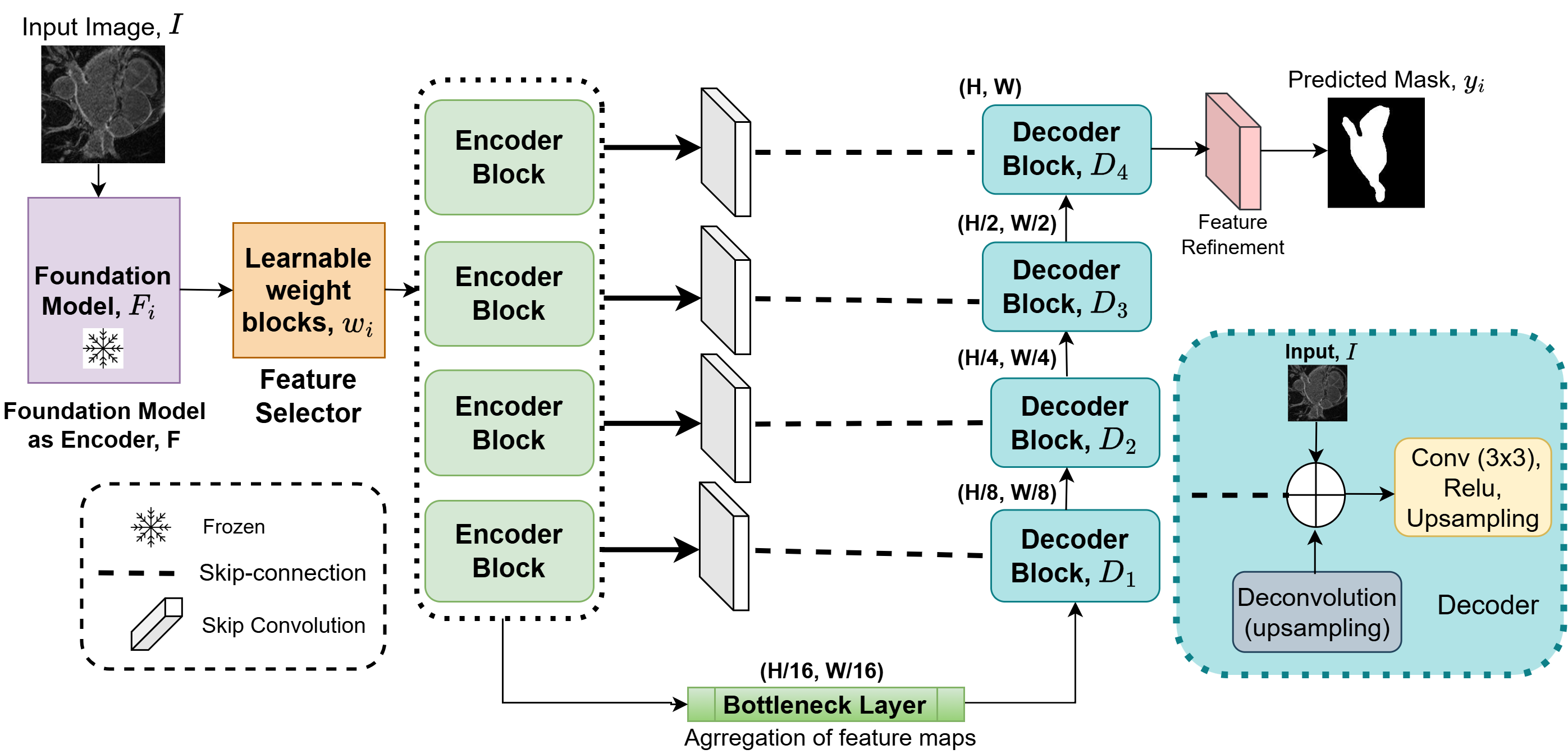 This study explores the robustness of vision foundation models for automated left atrium segmentation from cardiac MR images, addressing the critical challenge of limited annotated medical data in clinical settings. Natural domain foundation models were implemented with different decoder architectures and adaptation techniques to evaluate their segmentation performance on LGE-MRI data. As the lead researcher, I fine-tuned and benchmarked multiple vision transformer-based models.
Evaluating DINOv2 for Left Atrium Segmentation from MRI Images
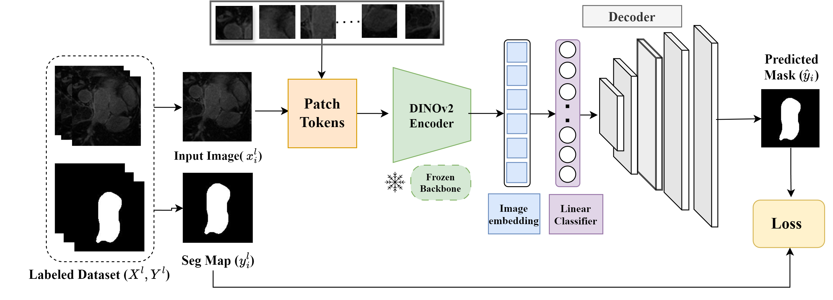 Accurate left atrium segmentation is crucial for diagnosing and planning treatments for atrial fibrillation. We evaluated the out-of-the-box performance of DINOv2, a self-supervised vision transformer, for segmenting the left atrium from MRI images. With a mean Dice score of 87.1%, DINOv2 outperformed baseline models, demonstrating its robustness even with limited data and minimal fine-tuning. This highlights DINOv2’s potential for broader applications in medical imaging.
Analysis of Non-rigid Registration Techniques
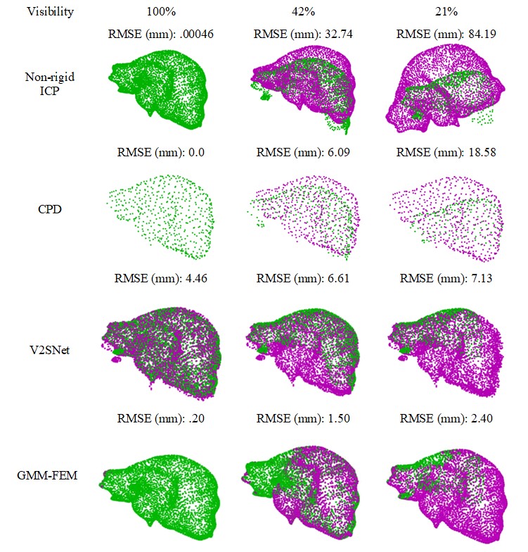 Non-rigid surface-based soft tissue registration is vital for surgical navigation, enabling the integration of pre-operative and intra-operative images for real-time visualization in a common coordinate system. Despite its potential, challenges like complex surface structures and degrees of freedom hinder widespread adoption. This study compares several open-source liver registration algorithms, highlighting the Gaussian Mixture Model-Finite Element Model (GMM-FEM) as the most robust, with consistently lower post-registration errors under reduced visibility and increased surface deformation. This method offers a promising solution for improving surgical navigation accuracy.